-
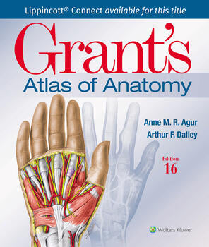
Illustrations drawn from real specimens, presented in surface-to-deep dissection sequence, set Grant's Atlas of Anatomy apart as the most accurate illustrated reference available for learning human anatomy and referencing in dissection lab. A recent edition featured re-colourisation of the original Grant's Atlas images from high-resolution scans, also adding a new level of organ luminosity and tissue transparency. The dissection illustrations are supported by descriptive text legends with clinical insights, summary tables, orientation and schematic drawings, and medical imaging.
- Renowned, high-resolution, dynamically colored illustrations organised in dissection sequence enable the formation of 3D constructs for each body region and provide detailed, realistic reference during dissection.
- Tables detail muscles, vessels, and other anatomic information in an easy-to-use format ideal for review and study.
- Enhanced medical imaging includes more than 100 clinically significant MRIs, CT images, ultrasound scans, and corresponding orientation drawings to help students confidently apply the laboratory experience to clinical rotations.
- Colour schematic illustrations reinforce the relationships of structures and anatomical concepts in vibrant detail.
- Fiction
- Children's and Young Adults Fiction
- Classic Fiction
- Contemporary Fiction
- Crime Fiction
- Graphic Novels
- LGBTIQA+
- New Zealand Fiction
- Poetry
- Children's and Young Adults Fiction
- Children's and Young Adult Fiction
- Pasifika Children's and Young Adult Fiction
- Picture Books
- Māori Children's and Young Adult Fiction
- New Zealand Children's and Young Adult Fiction
- New Zealand Picture Books
- Contemporary Fiction
- Fantasy
- Historical Fiction
- Horror and Ghost Stories
- Science Fiction
- Short Stories
- New Zealand Fiction
- New Zealand Classic Fiction
- New Zealand Crime and Thrillers
- New Zealand Fantasy and Sci-Fi
- New Zealand Graphic Novels and Manga
- New Zealand Historical Fiction
- New Zealand Horror and Ghost Stories
- New Zealand Short Stories
- Non-Fiction
- Arts and Artists
- Business and Law
- Health and Wellness
- Humanities
- Language and Education
- Lifestyle Books
- Māori Studies Books
- New Zealand Non-Fiction
- Pasifika Books
- Reference
- Science and Technology
- Social Sciences
- LGBTIQA+ Non-Fiction
- Medicine
- Veterinary Medicine
- Nursing
- Arts and Artists
- Architecture
- Art and Artists
- Dance
- Design and Graphic Design
- Film Studies
- Media Studies
- Music
- Photography
- Theatre Studies
- Business and Law
- Accounting and Finance
- Business Communication Studies
- Business Information Systems
- Business Statistics
- Careers Advice
- Economics
- International Business
- Investment
- Law
- Management
- New Zealand Business Studies
- New Zealand Law
- Tourism and Hospitality
- Health and Wellness
- Diet and Nutrition
- Health and Wellness
- Mind Body Spirit
- Self-help and Personal Development
- Humanities
- Ancient History
- Ancient Languages
- Anthropology and Archaeology
- Disability Studies
- Gender Studies
- History
- Philosophy
- Politics
- Religion and Spirituality
- Language and Education
- Education
- ESOL and ELT
- Language and Literacy
- Language Teaching and Learning
- Languages Chinese
- Languages French
- Languages German
- Languages Italian
- Languages Japanese
- Languages Korean
- Languages Spanish
- Library Studies
- Linguistics
- Literary Studies
- Lifestyle Books
- Biography and Memoirs
- Crafts and Hobbies
- Food and Drink
- Gardening
- Humour
- Popular Science
- Sport and Outdoor Recreation
- Travel Writing and Guides
- Māori Studies Books
- Disputed Land Whenua tautohetohe
- Māori Arts and Artists
- Māori Biography and Memoirs Haurongo
- Māori Crafts Mahi ā-ringa
- Māori Culture Mātauranga Māori
- Māori Education Mātauranga
- Māori Fiction and Poetry
- Māori Film and Media Studies
- Māori Food And Drink Kai me te inu
- Māori Health and Wellness Hauora
- Māori History Kōrero nehe
- Māori Indigenous Knowledge Mātauranga Māori
- Māori Mythology Pūrākau
- Māori Politics Tōrangapū
- Māori Religion Whakapono
- Matariki
- Te Reo Māori
- Treaty of Waitangi Tiriti o Waitangi
- New Zealand Non-Fiction
- New Zealand Architecture
- New Zealand Arts and Artists
- New Zealand Biography and Memoirs
- New Zealand Education
- New Zealand Environment and Sustainability
- New Zealand Film and Media Studies
- New Zealand Food and Drink
- New Zealand Gardening
- New Zealand Health and Wellness
- New Zealand History
- New Zealand Natural History
- New Zealand Photography
- New Zealand Politics
- New Zealand Social Services Welfare and Criminology
- New Zealand Sociology
- New Zealand Sport and Outdoors
- New Zealand Travel Writing and Guides
- Religion in New Zealand
- Pasifika Books
- Pacific Arts Crafts and Architecture
- Pacific Biography and Memoirs
- Pacific Culture and Indigenous Knowledge
- Pacific Education
- Pacific Fiction and Poetry
- Pacific Food and Drink
- Pacific Health and Wellness
- Pacific History and Politics
- Pacific Law
- Pacific Natural History
- Pacific Photography Film and Media Studies
- Reference
- Children's and Young Adult Reference
- Dictionaries
- General Reference
- Information Sciences
- New Zealand Children's Reference
- Research Methods
- Social Sciences
- Counselling and Therapy
- Psychology
- Social Services Welfare and Criminology
- Social Work
- Sociology
- Medicine
- Administration and Ethics
- Anaesthesiology
- Anatomy and Physiology
- Cardiology
- Clinical and Internal medicine
- Complementary Health
- Dentistry
- Dermatology
- Emergency medicine
- Endocrinology
- Family medicine
- Immunology and Infectious Diseases
- Medical Genetics
- Medical Study Guides
- Neurodiversity
- Neurology
- Obstetrics and Gynecology
- Oncology
- Ophthalmology
- Orthopaedics
- Paediatrics
- Pain Medicine
- Paramedic Studies
- Pathology
- Pharmacology
- Physical medicine and rehabilitation
- Psychiatry and Mental Health
- Public Health Medicine
- Radiology
- Respiratory Medicine
- Sexual Health Medicine
- Speech and Language Disorders
- Sport and Exercise Medicine
- Surgery
- Trans and Non-binary Health
- Urology
- Nursing
- Community Nursing
- Critical Care Nursing
- Geriatric Nursing
- Medical Surgical Nursing
- Mental Health Nursing
- Midwifery
- Nurse Leadership and Education
- Nursing Fundamentals and Skills
- Nursing Pharmacology
- Nursing Study Guides
- Occupational Therapy
- Paediatric Nursing
- Palliatve Care Nursing
- Textbooks
- ACG
- AUT
- College of Natural Health and Homeopathy
- Northtec
- South Pacific College of Natural Medicine
- Toi Ohomai Institute of Technology
- Unitec Institute of Technology
- University of Auckland
- Wintec Languages
- Stationery & Gifts
- Pens
- Pencils
- Highlighters and Markers
- Exercise Books and Pads
- Planning and Revision
- Art and Craft
- Filing and Storage
- Maths Equipment
- Wrapping
- Gifts and Giftware
- Art and Craft
- Art Pads
- Drawing Supplies
- Glue
- Painting Supplies
- Pins
- Printing Paper and Card
- Scissors and Knives
- Tape
- Visual Diaries
- Filing and Storage
- Binding Materials
- Clipboards
- Clips and Rings
- Dividers
- Document Wallets
- Expanding Files
- Hole Punches
- Pencil Cases and Pen Holders
- Presentation Folders
- Ringbinders
- Rubber Bands
- Staplers and Staples
- Gifts and Giftware
- Bookmarks
- Chocolate
- Colouring Books
- Journals
- Mugs
- Posters
- Playing Cards
- Puzzles
- Tarot Cards
- Tea
- Tote Bags
- Promotions
- Clearance Corner
- 15% off over 300+ Summer Reads
- 15% Off: Fourth Wing & Iron Flame
- 20% Off: Onyx Storm Pre-Orders
- 20% Off: Select Kids & YA Books
- Books of the Month - January
- Up to 20% Off: Virtual Book Club
We've sent you an email with a link to update your password.
Login
Reset your password
We will send you an email to reset your password.


